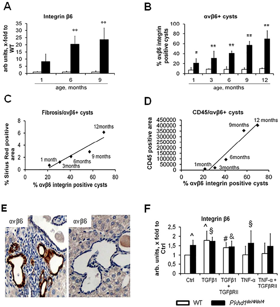
Figure 4. αvβ6 integrin expression correlates with portal fibrosis and peribiliary accumulation of CD45+ cells in Pkhd1del4/del4 mice.
A. In contrast with WT mice, Pkhd1del4/del4 mice showed a progressive increase in β6 gene expression in whole liver, which was greater than 25-fold at 6 months and nearly 35-fold at 9 months (n=3 for each age). B. Similarly, αvβ6 integrin expression on cyst epithelia assessed by immunohistochemistry increased progressively, reaching nearly the 70% of biliary structures at 12 months (n=3/5 for each age). C–D. Immunohistochemical expression of αvβ6 strongly correlated both with portal fibrosis (r=0.94, p<0.02) and with the CD45+ portal cell infiltrate (r=0.97, p<0.01). E. In any given specimen, αvβ6 integrin was typically higher in biliary cysts surrounded by a dense inflammatory infiltrate (immunoperoxidase for αvβ6, representative sample, 6 months, M= 400×). F. TGFβ1 and TNFα stimulate gene expression of β6 integrin in Pkhd1del4/del4 cholangiocytes. In Pkhd1del4/del4 cholangiocytes, β6 mRNA was significantly increased in basal conditions with respect to WT. In both WT and Pkhd1del4/del4 cholangiocytes, β6 expression was further and significantly increased after TGFβ1. Unlike TGFβ1, TNFα significantly increased β6 mRNA expression only in Pkhd1del4/del4 cholangiocytes. TGFβRII blockade abolished the TGFβ1 effects on β6 mRNA expression in both Pkhd1del4/del4 and WT cholangiocytes, in contrast to TNFα whose effects on β6 were unaffected by TGFβRII antagonism (n=6/10 for each experimental condition, cultured cholangiocytes derived from mice of 3 months of age). °°p<0.01 vs WT (1 month), *p<0.05 vs WT (same age), **p<0.01 vs WT (same age), ^p<0.05 vs WT Ctrl, §p<0.05 vs Pkhd1del4/del4 Ctrl, #p<0.05 vs WT TGFβ1-treated, &p<0.05 vs Pkhd1del4/del4 TGFβ1-treated.

