. 2016 Feb 16;10(1):99–110. doi: 10.4184/asj.2016.10.1.99

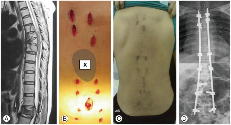
Fig. 1. Case 22: A 58-year-old woman with lung cancer presented with pathological fracture of L2 with cauda equine syndrome. One month prior to the presentation, she had radiotherapy to the T6–T7 region for pathological fractures. (A) MRI showed pathological fractures of T6, T7, and L2 with spinal canal stenosis at L2 lead to cauda equine syndrome. (B) Intraoperative photo showed a multiple small stab wounds for percutaneous pedicle screws insertion. A minimally invasive direct decompression was performed at L2 region with cement augmentation for L2 fracture. The minimally invasive stabilization (MISt) construct spared the radiated zone (X). (C) Postoperative 2 weeks, the patient was able to sit up and mobilize with minimal pain. (D) Postoperative radiograph showed Long Construct MISt from T3-L5 spanning 15 spinal segments.

Fig. 1