. 2015 Nov 27;4:e09600. doi: 10.7554/eLife.09600

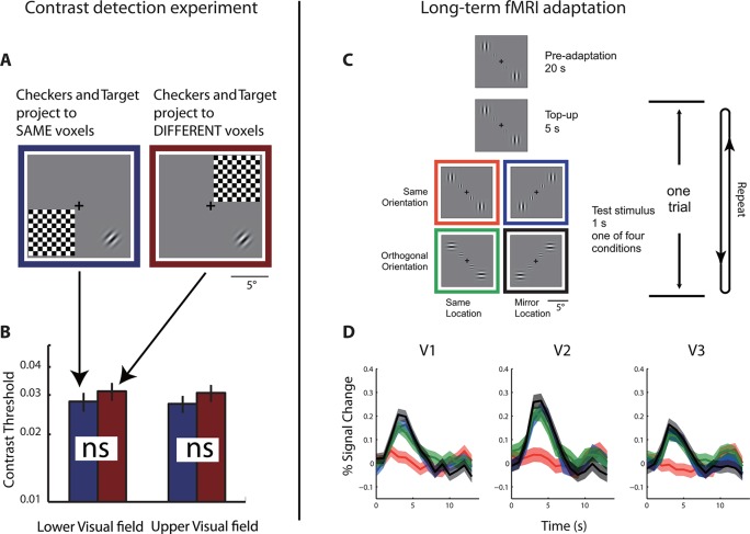
Figure 2. Behavioral and physiological evidence of independence between the co-localized neural populations.

(A) Schematic of the psychophysical experiment of contrast detection with a contralateral mask. The 45° target Gabor patch was presented in either the lower-right (shown) or upper-left quadrant. A high-contrast flickering checkerboard mask was presented at the mirrored location from the target quadrant, across either the vertical or horizontal meridian. S was to identify which one of the two temporal intervals the target appeared in. (B) Contrast detection thresholds were essentially the same for the two mask-target arrangements. Error bars denote ± SE across blocks. (C) Design of the fMRI adaptation experiment. Each block of trials was preceded with 20 s of pre-adaptation. Each trial began with a 5 s presentation of the adapting stimulus (‘top-up’ adaptation), followed by one of the four test stimuli. Relative to the adapting stimulus, the test stimulus could either be at the same or mirrored location, and could have either the same or orthogonal orientation. Attention was controlled with a demanding central fixation task. (D) Time courses of fMRI BOLD responses in V1-V3 to the four test conditions. Shaded error band denote ± SE across trials. The responses evoked by the test stimuli presented at the mirrored location relative to the adaptor, regardless of orientation, did not differ significantly from those evoked by the test stimuli at the same location as the adaptor but with orthogonal orientation.

DOI: http://dx.doi.org/10.7554/eLife.09600.005

Figure 2.

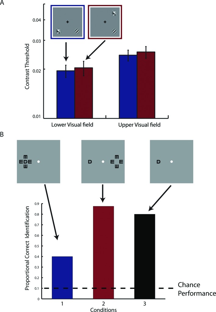
Figure 2—figure supplement 1. Results from supplementary psychophysical experiments implicating two groups of non-interacting neurons.

Figure 2—figure supplement 1.

(A) Contrast detection task with a contralateral noise mask. The target Gabor patch was presented in either the lower-right (shown) or upper-left quadrant. An isotropic noise mask of the same spatial frequency band as the target was presented at the mirrored location of the target across either the vertical or horizontal meridian. In the cross-vertical-meridian arrangement (blue), but not in the cross-horizontal-meridian arrangement (red), both the target and the mask projected to the same cortical locations in V1-V3 of the achiasmic subject S. S was to identify which one of the two temporal intervals contained the target. Contrast detection thresholds were essentially identical for the two mask positions. Error bars denote ± SE across different blocks. (B) Letter identification task: S’s task was to identify the target (center) letter presented at an eccentricity of 10 deg and flanked with the 4 tumbling E’s. The target letter was randomly drawn from the set of 10 letters (C, D, H, K, N, O, R, V, S and Z). The stimuli appeared for 150 ms at a given location on the CRT monitor. There were three conditions: (1) the flankers and target on the same side, (2) the flankers and target on symmetrically opposite sides across the vertical meridian, and (3) target only. When the flankers and the target were on the same side (Condition 1), letter identification performance was severely impaired by the flankers -- the well-known crowding effect. When the flankers and target were on the opposite sides (Condition 2), even though the cortical representations of the flankers and target were equally close as in Condition 1, no crowding effect was found, and performance was identical to the target-only condition. These behavioral experiments show that even though the cortical representations of left and right visual fields overlap in V1-V3 in achiasma, the neuronal populations that represent these visual fields do not interact functionally.