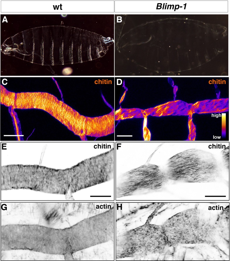
Figure 4. Taenidial folds, F-actin bundles, and chitin levels in Blimp-1 mutant embryos.
(A-B) Cuticle preparations of wild-type (A) and Blimp-1 (B) embryos visualised under dark field. In the Blimp-1 mutant embryo, the cuticle and the denticle belts, chitin structures at the epidermis, are faint when compared to wild-type preparation. (C-D) Stage 17 Blimp-1 heterozygous (control, C) and Blimp-1 homozygous mutant (D) embryos stained with fluostain to label chitin structures. After acquisition in the same conditions, the images are converted into colour-coded LUTs in which different levels of fluorescent signals are matched with different colours. The colour code is shown on the lower right hand of panel D. While in the control DT mostly red and yellow stains are observed, in the Blimp-1 mutant DT there are mostly purple and red stains, indicating that the fluorescent signal level of fluostain is lower in the Blimp-1 mutant DT. Scale bars = 10 μm. (E-H) Wild-type (E, G) and Blimp-1 mutant embryos (F, H) stained with fluostain (E, F) to label taenidial folds and phalloidin (G, H) to label F-actin bundles. Both the taenidial folds and F-actin bundles run perpendicular to the tube axis in wild-type embryos (E, G) while in most of the Blimp-1 mutants F-actin bundles fail to form (H) and taenidial folds run parallel to the tube axis (F). The images are single stacks of confocal sections. Scale bars = 10 μm.

