. 2016 Feb 15;5:e10696. doi: 10.7554/eLife.10696

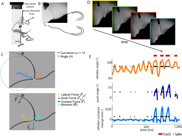
Figure 1. Electrophysiological recording from single primary whisker units in awake, head-fixed mice and simultaneous measurement of whisker kinematics/mechanics. .

(A) Schematic of the preparation, showing a tungsten microelectrode array implanted into the trigeminal ganglion of a head-fixed mouse, whilst a metal pole is presented in one of a range of locations (arrows). Before the start of each trial, the pole was moved to a randomly selected, rostro-caudal location. During this time, the whiskers were out of range of the pole. At the start of the trial, the pole was rapidly raised into the whisker field, leading to whisker-pole touch. Whisker movement and whisker-pole interactions were filmed with a high-speed camera. (B, C) Kinematic (whisker angle θ) and mechanical (whisker curvature κ, moment M, axial force Fax and lateral force Flat) variables were measured for the principal whisker in each video frame. When a whisker pushes against an object during protraction (as in panel D, red and cyan frames), curvature increases; when it pushes against an object during retraction (as in panels B and C), it decreases. (D) Individual video frames during free whisking (yellow and green) and whisker-pole touch (red and cyan) with tracker solutions for the target whisker (the principal whisker for the recorded unit, panel E) superimposed (coloured curve segments). (E) Time series of whisker angle, push angle and curvature change, together with simultaneously recorded spikes (black dots) and periods of whisker-pole contact (red bars). Coloured dots indicate times of correspondingly coloured frames in D.

DOI: http://dx.doi.org/10.7554/eLife.10696.003

Figure 1.

Figure 1—figure supplement 1. Electrophysiological recording from trigeminal primary neurons of awake, head-fixed mice. .

Figure 1—figure supplement 1.

Extracellular potential recorded from the same single unit during both anaesthetized and awake epochs. Spikes belonging to the cluster of the target unit are shown by black triangles. Inset shows overlay of all waveforms belonging to this cluster.
Figure 1—figure supplement 2. Computation of axial and lateral contact forces. .

Figure 1—figure supplement 2.

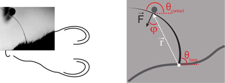
Axial (Fax) and lateral (Flat) force components at the whisker base were calculated, in each video frame where there were whisker-pole contacts, as follows (Pammer et al., 2013). First, the point of whisker-pole contact was located (Materials and methods). The direction of the force F was then calculated as the normal to the whisker tangent at the contact point (Pammer et al., 2013). Moment at the base M was calculated from the whisker curvature at the base (Materials and methods) and then the magnitude F of F was derived from the definition of moment:
F=Mrsin(φ)
where r is the magnitude of the lever arm vector r from whisker base to contact point, and φ is the angle between r and F. The components Fax and Flat were then found by projecting F onto the tangent and normal to the whisker at its base, respectively:
Fax=Fsin (θbaseθcontact),
Flat=Fcos (θbaseθcontact).
Here θbase is the angle between the tangent to the whisker at its base and the horizontal; θcontact is the angle between F and the horizontal.