. 2015 Dec 23;4:e12401. doi: 10.7554/eLife.12401

Figure 1. Pigment cell projections.

(A) Zebrafish and pearl danio. Right, melanophores and xanthophores (arrows) after epinephrine treatment to contract pigment granules. (B) Long projections by zebrafish aox5+ cells of xanthophore lineage (arrows) with membraneous vesicles (arrowhead, inset). (C) Zebrafish aox5+ cells were more likely to extend projections than pearl, especially during early stripe development [7–8 SSL (Parichy et al., 2009); species x stage, χ2=103.4, d.f.=4, p<0.0001; N=929, 1259 cells for zebrafish and pearl; projections per cell: χ2=45.3, d.f.=1, p<0.0001]. (D) In zebrafish, projections were often long and fast. Bars indicate median ± interquartile range (IQR). (E) Extension and retraction (arrow) and release of vesicle (arrowhead) in zebrafish but not pearl. Scale bars: 5 mm (A, left); 50 µm (A, right); 10 µm (B); 50 µm (E).

DOI: http://dx.doi.org/10.7554/eLife.12401.003

Figure 1.

Figure 1—figure supplement 1. Developing adult pigment patterns during peak stages of aox5+ fast projections in zebrafish.

Figure 1—figure supplement 1.

In zebrafish (7.5 SSL), melanophores are found along the horizontal myoseptum (e.g., outline and inset) and an interstripe region with pigmented xanthophores (inset, arrow) has started to develop. Vertical bars at left indicate approximate locations of stripes (black) and interstripes (orange). In pearl, fewer melanophores are present, the developing interstripe is indistinct and xanthophores have differentiated widely across the flank. Scale bar: 200 µm.

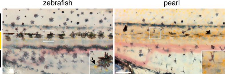
Figure 1—figure supplement 2. Zebrafish aox5+ cells extend fast projections independently of melanophores and iridophores.

Figure 1—figure supplement 2.

(A) aox5+ projections persisted in microphthalmia a (mitfa) mutants (Lister et al., 1999), lacking melanophores, though were somewhat less frequent than in wild-type (wt; χ2=7.4, d.f.=1, p<0.01). The incidence of aox5+ cells extending projections did not differ from wild-type in leucocyte tyrosine kinase (ltk) mutants (Lopes et al., 2008) that lack iridophores (p=0.1). (B) Left, excess interstripe melanophores were generated by repeated heat-shock induction of ubiquitous Kit ligand a (Kitlga) (Patterson and Parichy, 2013; Patterson et al., 2014) (F1,5=12.9, p<0.05; control, heat-shocked wild-type siblings lacking the transgene). Middle, Increased numbers of melanophores did not affect the frequency with which projections were extended by aox5+ cells (p=0.9). Right, Despite increased numbers of melanophores, there were no effects on interstripe xanthophore numbers (p=0.8), suggesting that aox5+ cells do not alter their differentiation in response to whether or not they are in the vicinity of melanophores.

Figure 1—figure supplement 3. Rare fast projections of zebrafish melanophores.

Figure 1—figure supplement 3.

(A) Incidences of projections by tyrp1b+ and aox5+ cells of melanophore and xanthophore lineages, respectively. (B and C) Examples of tyrp1b+ melanophores extending projections that were indistinguishable from those of aox5+ cells; labeling: membrane targeted mCherry (B) and GFP (C). (D) High resolution image of tyrp1b+ melanophore, illustrating abundant, short filopodial projections (arrowhead). Scale bars: 50 µm (B and C); 10 µm (D).