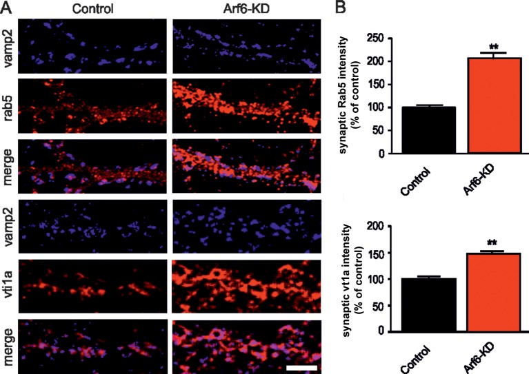
Figure 2. Increased expression of endosomal markers at Arf6-deficient synapses.
(A) Representative images of synapses from rat hippocampal neurons (17 DIV) transduced with either Arf6 shRNA (Arf6-KD) or an inactive mismatched version (Control) and immunostained with anti-Vamp2 (blue) and either anti-Rab5 or anti-Vti1A (red) antibodies. Scale bar, 5 µm. (B) Intensity values for Rab5 and Vti1A signal at VAMP2-positive puncta in control (black) and Arf6-silenced (red) synapses. Data are means ± SEM from 3 independent preparations. 500 synapses have been counted for each preparation. Statistical analysis was performed with the unpaired Student's t-test. **p<0.005 versus control.

