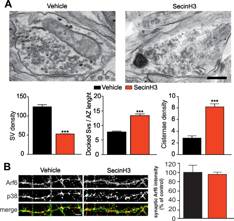
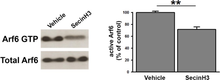
Figure 3. SecinH3 treatment phenocopies Arf6 silencing.
(A) Upper panels, representative electron micrographs of synaptic terminals from cultured hippocampal neurons (17 DIV) treated with either SecinH3 (30 µM) or DMSO (Vehicle). Lower panels, morphometric analysis of the density of SVs, docked SVs and cisternae in control (black) and SecinH3 treated (red) synapses. Data are means ± SEM from 3 independent preparations (n=75 for experimental group). Statistical analysis was performed with the unpaired Student’s t-test. ***p<0.001, versus respective control. Scale bar, 200 nm. (B) Left panels, representative images of synapses from rat hippocampal neurons (17 DIV) treated with SecinH3 (30 µM) or DMSO (Vehicle) and immunostained with anti-Arf6 and anti-synaptophysin (p38) antibodies. Scale bar, 5 µm. Right panel, intensity values for Arf6 signal at p38-positive puncta in control (black) and SecinH3-treated (red) synapses. Data are means ± SEM from 3 independent preparations. 300 synapses have been counted for each preparation.


