. 2015 Oct 27;4:e09946. doi: 10.7554/eLife.09946

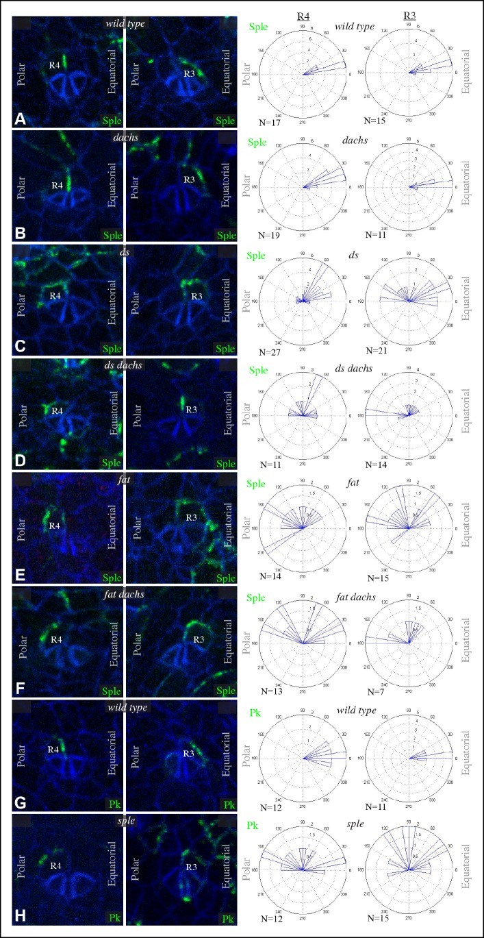
Figure 5. Sple and Pk localization in photoreceptor cells.

Localization of GFP:Sple (A–F) or GFP:Pk (G,H) in cells with expression in R4 or R3 photoreceptor cells in wild type (A,G), dGC13/ d210 (B), ds36D/ dsUA071 (C), dGC13 ds36D/ dGC13 dsUA071 (D), ft8/ ftG-rv (E), dGC13 ft8/ dGC13 ftG-rv (F), and sple1 (H) mutants. Rose plots summarize localization based on the indicated number (N) of examples, with equatorial to the right, polar to the left, and anterior (towards the morphogenetic furrow) at top.

DOI: http://dx.doi.org/10.7554/eLife.09946.010

Figure 5.

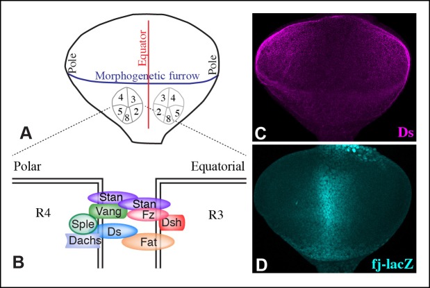
Figure 5—figure supplement 1. Polarity and gradients in eye discs.

Figure 5—figure supplement 1.

(A) Cartoon illustrating the arrangement and orientation of photoreceptor cells in eye discs at the 5-cell pre-cluster stage. (B) Schematic illustrating the subcellular localization of Ds-Fat and Fz PCP pathway components at the R4 and R3 interface. (C,D) Expression of Ds (magenta) and fj-lacZ (cyan) in a wild-type eye disc.
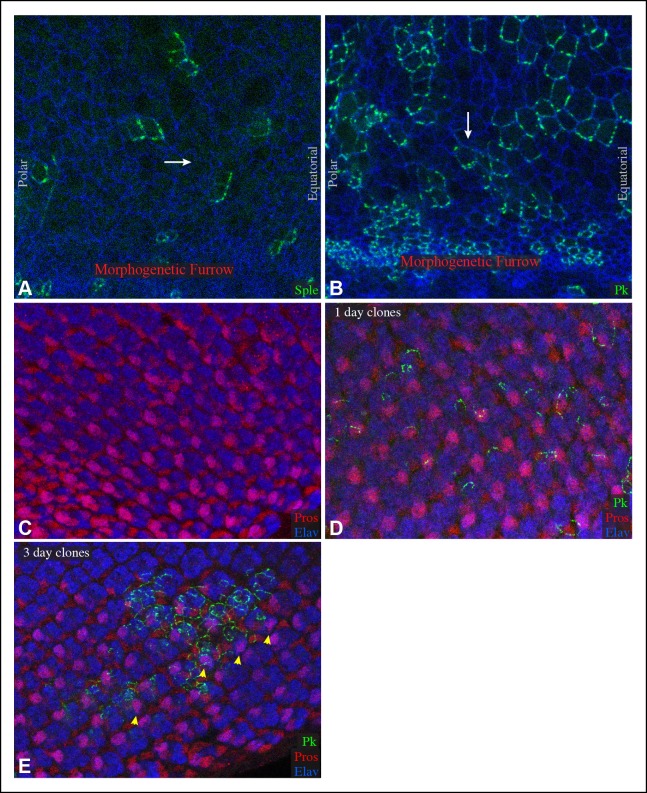
Figure 5—figure supplement 2. Sple and Pk polarity in front of the morphogenetic furrow, and influence of GFP:Pk on PCP.

Figure 5—figure supplement 2.

(A,B) Portions of eye imaginal discs in front of the morphogenetic furrow with clones of cells expressing GFP:Sple (A) or GFP:Pk (B) (green), stained for expression of E-cadherin (blue). The white arrows indicates general direction of Sple or Pk polarization. (C,D) Portions of eye imaginal discs stained for Elav (marks photoreceptor cells) and Prospero (Pros, overlap with Elav marks R7) and expressing GFP:Pk (green). (C) control disc with no GFP:Pk clones. (D) Disc with 1-day-old GFP:Pk clones. (E) Disc with 3-day-old GFP:Pk clones, yellow arrowheads highlight ommatidia with abnormal polarity.