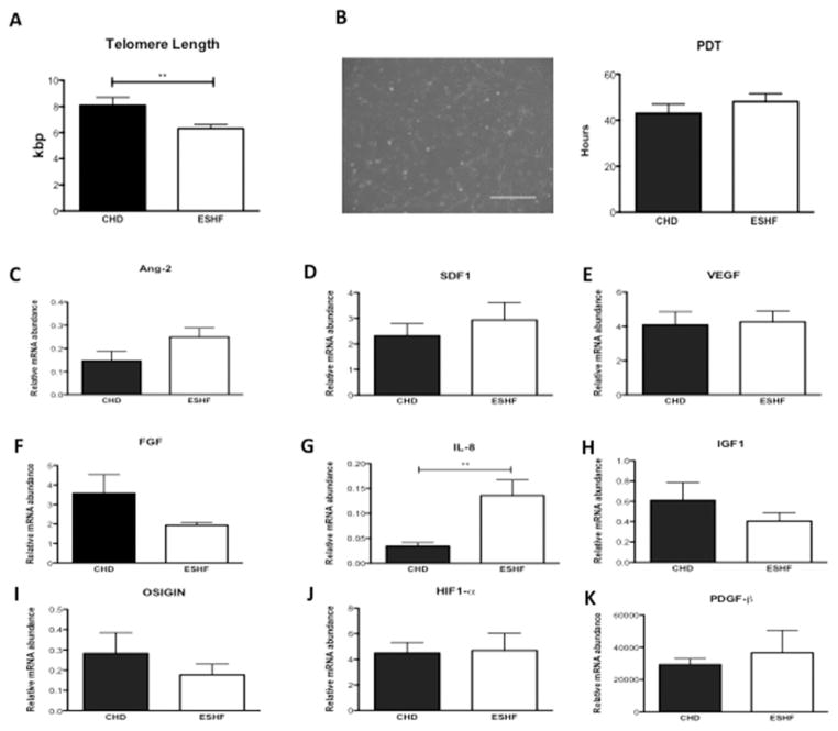
Fig 4.
Growth properties of end-stage heart failure (ESHF) versus congenital heart disease (CHD)-derived c-kit+ cardiac stem cells and in vivo paracrine factor expression. (A) Comparison of telomere length in c-kit+ cardiac stem cells isolated from CHD versus ESHF myocardium. (B) Representative phase-contrast microscopy image of c-kit+ cardiac stem cell morphology with quantification of population doubling time. (C–K) In vivo comparison of growth factors (CHD, n = 5 versus ESHF, n = 5 for each): (C) angiopoietin 2 (Ang-2), (D) stromal-derived factor 1 (SDF-1), (E) vascular endothelial growth factor (VEGF), (F) fibroblast growth factor (FGF), (G) interleukin 8 (IL-8), (H) insulin growth factor 1 (IGF-1), (I) oxidative stress-induced growth inhibitor 1 (OSIGIN), (J) hypoxia-inducible factor 1 (HIF-1α), and (K) platelet-derived growth factor (PDGF-β). **p < 0.01.

