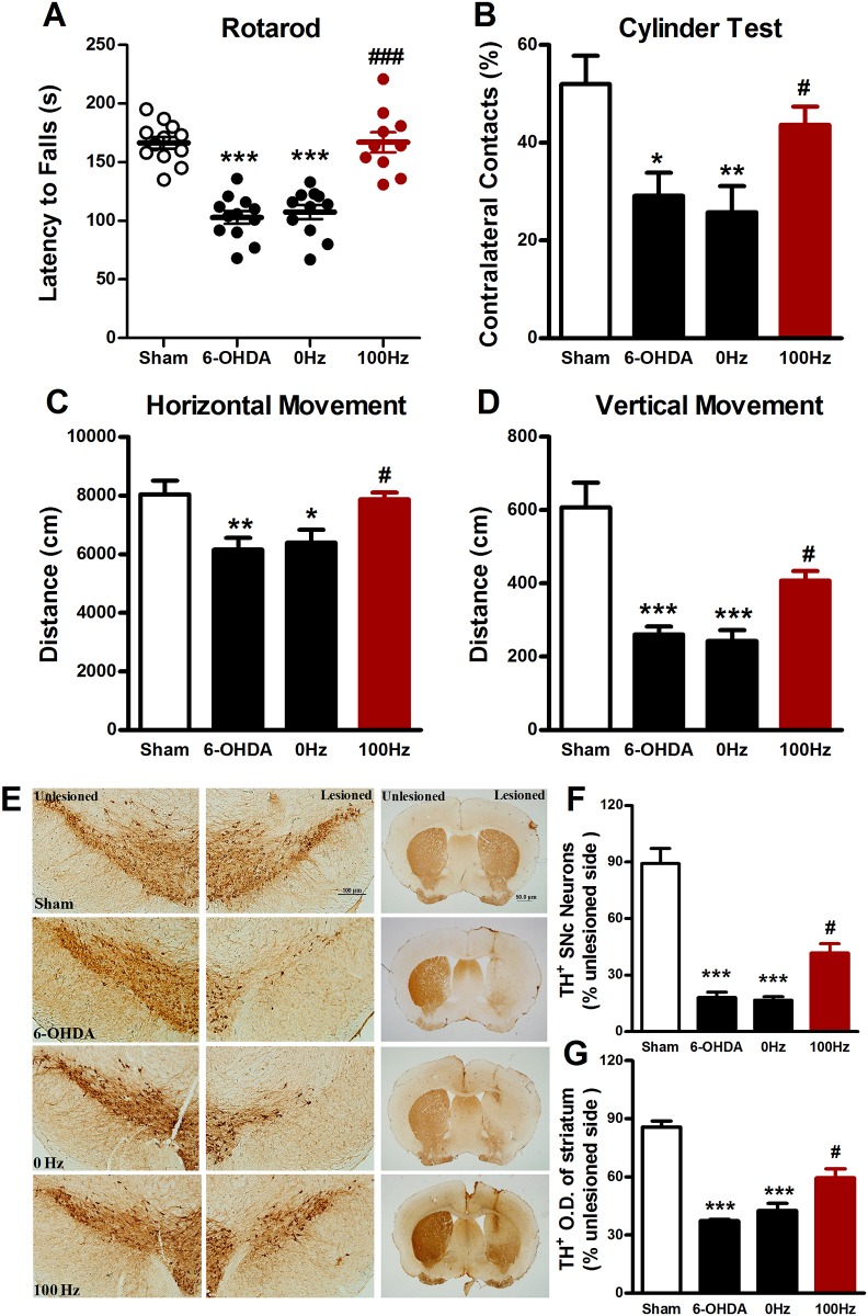
Fig 3. Four weeks’ EA stimulation exerted the behavioral improvement and neuroprotective effects on 6-OHDA-lesioned mice.
(A) Rotarod test. (B) Cylinder test. (C) Horizontal locomotor distance. (D) Vertical locomotor distance. (F) Immunohistochemistrical staining for TH positive dopaminergic neurons in the SNpc (scale bar: 100 μm) and fibers in the striatum (scale bar: 50 μm). (G) The percentage of TH positive dopaminergic neurons in the SNpc (H) The percentage of average optical density of TH positive dopaminergic fibers in the striatum. The data were expressed as means ± S.E.M. n = 10–12 per group for the behavior tests; n = 5 per group for the immunohistochemistrical experiments. The values were expressed as means ± SEM. * P <0.05; ** P < 0.01; *** P <0.001 vs. Sham group. # P <0.05; ### P <0.001 vs. 0 Hz group.

