Figure 12.
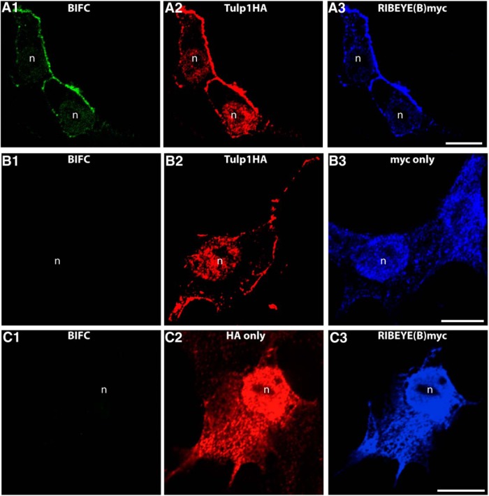
The indicated BIFC constructs were transfected into HEK293T cells and analyzed for fluorescence complementation (BIFC signal) in the green channel (mVenus). Expression of the separate plasmids was verified by immunolabeling with antibodies against the HA-tag (red channel) and with antibodies against myc-tag (far red channel, blue), exactly as previously described (Ritter et al., 2011). Fluorescence complementation was only observed if Tulp1 was cotransfected with RIBEYE(B) (A). Only in this case, we observed a BIFC signal (A1). If Tulp1 was transfected with empty control vectors (B), no fluorescence complementation was observed (B1). Similarly, if RIBEYE(B) was transfected with empty control plasmid (C), no BIFC signal was generated (C1). n = 4 experiments. n, nucleus of a transfected HEK293T cell. Scale bars, A–C, 5 μm.

