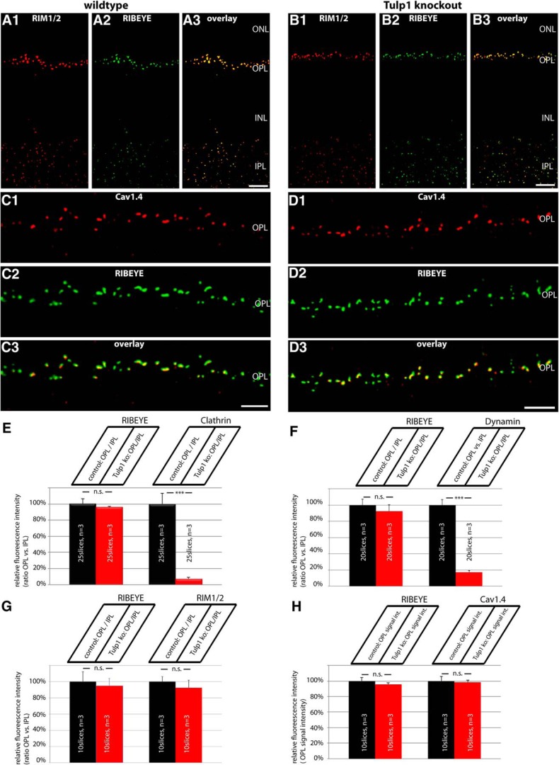
Figure 5.
A–D, Low-magnification micrographs of 0.5-μm-thin resin sections from Tulp1 knock-out (B, D) retinas and control (A, C) retinas that were immunolabeled with rabbit polyclonal antibodies against RIM1/2 and mouse monoclonal antibodies against RIBEYE in A, B and mouse monoclonal antibodies against Cav1.4 and rabbit polyclonal antibodies against RIBEYE in C, D. In both wild-type and Tulp1 knock-out mice, both RIM1/2 and Cav1.4 were strongly expressed in the OPL. In the OPL, where photoreceptor synapses are located, both RIM1/2 and Cav1.4 immunosignals were highly enriched at the synaptic ribbons, as previously described (Wahl et al., 2013). In contrast to the endocytic proteins of the periactive zone, the active zone proteins RIM1/2 and Cav1.4 remained unchanged in the OPL of Tulp1 knock-out mice. For high-magnification analyses of single photoreceptor ribbon complexes, see Figure 6. ONL, Outer nuclear layer; INL, inner nuclear layer; OPL, outer plexiform layer; IPL, inner plexiform layer. Scale bars: A, B, 15 μm; C, D, 5 μm. E–H, The expression of dynamin, clathrin heavy chain, and RIM1/2 at photoreceptor ribbon synapses in the OPL was quantified and related to the IPL to correct for possible differences in section thickness (E–G). Only in H (quantification of Cav1.4), absolute fluorescence numbers were given for the OPL (with the control animals set to 100%) because Cav1.4 is not expressed in significant amounts in the IPL (Grabner et al., 2015) (see also Materials and Methods). Quantification confirmed the loss of endocytic periactive zone proteins at photoreceptor synapses of Tulp1 knock-out mice, whereas the distribution of active zone proteins remains unaltered in Tulp1 knock-out mice. n.s., Nonsignificant. Error bars indicate SEM. ***p < 0.001.

