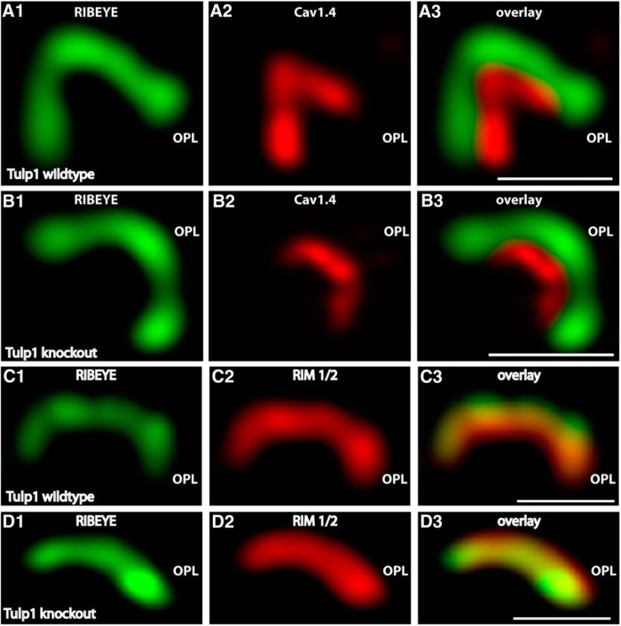
Figure 6.
A, B, Typical examples of single synaptic ribbon complexes (side views) of rod photoreceptor synapses double-immunolabeled with mouse antibodies against RIBEYE (B)/CtBP2 (A, B) and rabbit polyclonal antibodies against the pore-forming α1-subunit of Cav1.4 (A, B) in Tulp1 knock-out mice (B) and littermate control mice (A). Representative results obtained from four animals (each genotype) of four litters (three experiments from each wild-type and knock-out animal). C, D, Single synaptic ribbon complexes (side views) of rod photoreceptor synapses double-immunolabeled with mouse monoclonal antibodies against RIBEYE (C, D) and rabbit polyclonal antibodies against RIM1/2 (C, D) in Tulp1 knock-out mice (D) and littermate control mice (C). Three independent experiments from three animals of each genotype (littermate controls). In contrast to the endocytic proteins in the periactive zone, the localization of the active zone proteins Cav1.4 (A, B) and RIM1/2 (C, D) remained unchanged in the Tulp1 knock-out mice. All images were obtained by SR-SIM. OPL, Outer plexiform layer. Scale bars, 1 μm.

