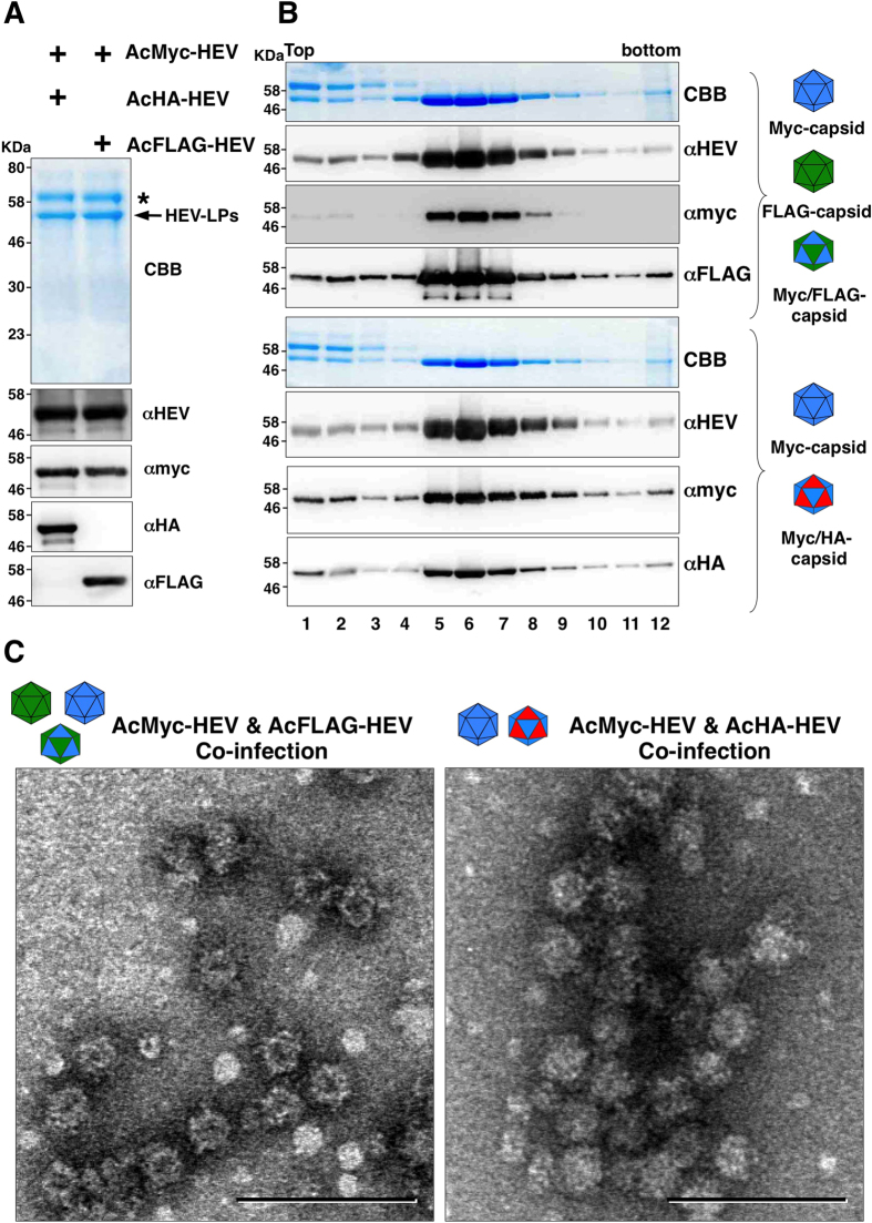
Figure 3. Production of chimeric HEV-LP possessing divalent foreign epitopes.
(A) CBB staining and immunoblotting of the culture supernatants of Tn5 cells infected with AcMyc-HEV (blue) together with either AcHA-HEV (red) or AcFLAG-HEV (green). Asterisk and arrow in the CBB staining gel indicate bovine serum albumin and recombinant HEV capsids, respectively. (B) Sucrose density gradient fractionation of the culture supernatants of cells co-infected with the recombinant viruses. Putative recombinant and chimeric HEV-LP were indicated in the left of the panels. Each fractions were subjected to CBB staining and immunoblotting after SDS-PAGE. (C) Electron microscopic observation of the middle fractions of the sucrose density gradient. HEV-LP were imaged by negative staining. Bars indicate 100 nm.

