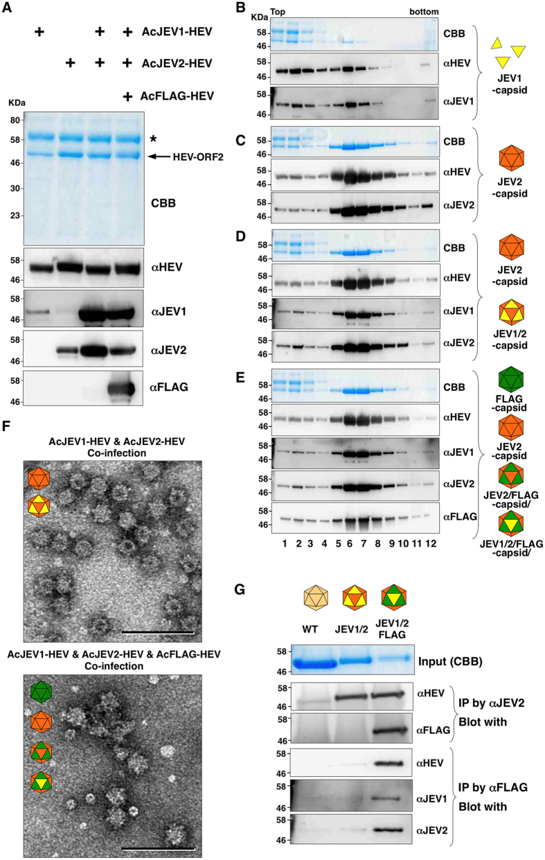
Figure 5. Production of chimeric HEV-LP bearing JEV neutralizing epitopes.
(A) CBB staining and immunoblotting of the culture supernatants of Tn5 cells infected with various combination of AcJEV1-HEV (yellow), AcJEV2-HEV (orange) and AcFLAG-HEV (green). Asterisk and arrow in the CBB staining gel indicate bovine serum albumin and recombinant HEV capsids, respectively. (B~E) Sucrose density gradient fractionation of the culture supernatants of cells infected with either AcJEV1-HEV (B) or AcJEV2-HEV (C), co-infected with AcJEV1-HEV and AcJEV2-HEV (D), or co-infected with AcJEV1-HEV, AcJEV2-HEV, and AcFLAG-HEV (E). Putative recombinant and chimeric HEV-LP were indicated in the left of the panels. Each fractions were subjected to CBB staining and immunoblotting after SDS-PAGE. (F) Electron microscopic observation of the middle fractions of the sucrose density gradient. HEV-LP were imaged by negative staining. Bars indicate 100 nm. (G) Incorporation of multiple recombinant capsids into HEV-LP. HEV-LP recovered from insect cells infected with AcHEV (lane 1), co-infected with AcJEV1-HEV and AcJEV2-HEV (lane 2), and co-infected with AcJEV1-HEV, AcJEV2-HEV, and AcFLAG-HEV (lane 3) were immunoprecipitated by either anti-JEV2 or -FLAG antibody and the immunoprecipitates were further examined by immunoblotting by indicated antibodies. In put of each HEV-LP stained by CBB is indicated on the top.

