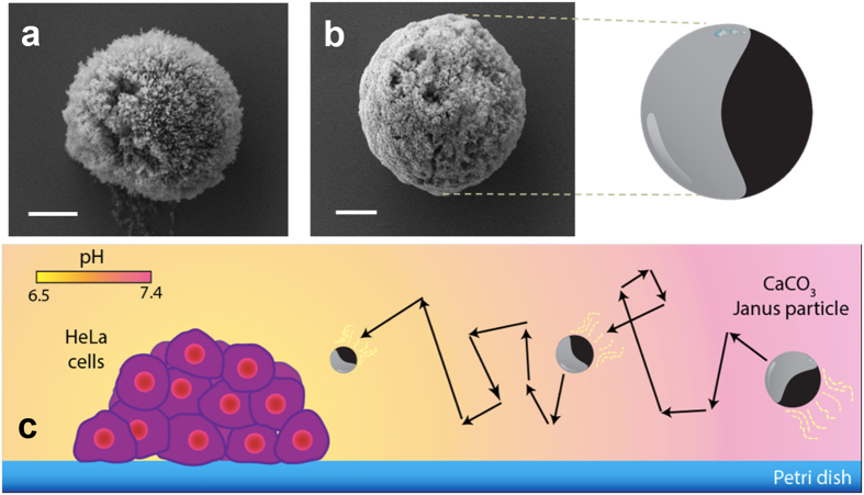
Figure 1. Morphological characterization of CaCO3 Janus particles.
SEM images of bare CaCO3 (a) and CaCO3 Janus particles, with a half of its surface covered with cobalt thin layer, along with the schematic from the CaCO3 Janus particles (b). Schematic of CaCO3 particles moving in acidic environment in situ generated by HeLa cells (c). Scale bar = 2 μm.

