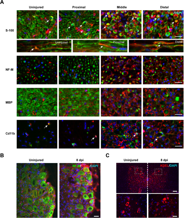
Figure 2. ER stress responses after sciatic nerve crush.
Wild-type mice were crush injured in the right sciatic nerve and sham operated in the left sciatic nerve. After 8 days, nerves were removed for histological analysis. (A) KDEL staining (red) was performed using indirect immunofluorescence and co-stained with S100 (Schwann cells), NF-M (axons), MBP (myelin), and a Cd11b (macrophages) in green. Cell nuclei were counter stained using DAPI (blue). Co-localization is denoted using white arrows and S100 negative KDEL positive cells with asterisk. Scale bar: 20 μm. (B) DRGs and were collected from animals described in A and KDEL staining (red) was performed together with the neuronal marker NF-M (green). Scale bars: 100 μm. (C) Thoracic spinal cord from animals described in A was analyzed for KDEL staining (red). Scale bars: 100 μm (low magnification) and 40 μm (high magnification).

