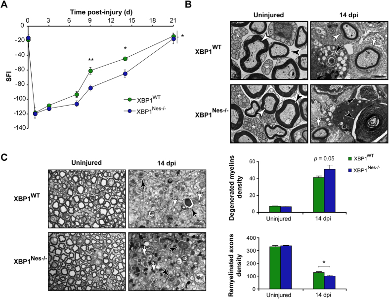
Figure 3. XBP1 deficiency decreases myelin removal, axonal regeneration and locomotor recovery after sciatic nerve injury.
(A) XBP1WT and XBP1Nes−/− mice were crush injured in the right sciatic nerve and sham operated in the left sciatic nerve. Locomotor recovery was evaluated using the SFI analysis before (0 day) and at indicated time points. (B) Electron microscopy of uninjured and distal segments of 14 dpi from XBP1WT and XBP1Nes−/− mice. White arrowheads indicate intact myelinated axons, black arrowheads, intact myelins, and asterisks unmyelinated axons. Black arrows point to degenerated myelins and white arrows, to remyelinated axons. Scale bar: 2 μm. (C) Transversal semi-thin sections of sciatic nerves stained with toluidine blue from uninjured and at 14 dpi XBP1WT and XBP1Nes−/− mice. Sections were obtained 3 mm distal to crush segment. Black and white arrows indicate demyelinated and regenerated fibers, respectively (left panel). Scale bar: 10 μm. Quantification of degenerated myelins and remyelinated axons density is presented (right panel). Data are shown as mean ± S.E.M.; *p < 0.05; **p < 0.01. SFI data were analyzed by repeated measures ANOVA followed by Bonferroni’s post hoc test (n = 7 animals per group). Morphological data was analyzed at each time point by student’s t-test (n = 3 animals per group).

