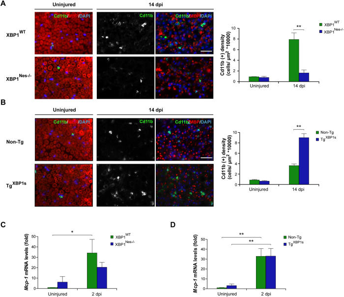
Figure 6. XBP1 expression in the nervous system enhances macrophage infiltration in injured sciatic nerves.
(A) Sciatic nerves from XBP1Nes−/− and XBP1WT littermates were processed for immunofluorescence from uninjured conditions and at 14 dpi distal sciatic nerves were analyzed for Cd11b (green) to evaluate macrophages and MBP (red) to stain myelin sheaths. Nuclei were counterstained using DAPI (blue, left panel). The staining density for Cd11b was quantified at 14 dpi in XBP1Nes−/− and XBP1WT mice (right panel). (B) TgXBP1s and non-Tg sciatic nerves were analyzed as described in A. Mcp-1 expression was analyzed in sciatic nerves of XBP1Nes−/− and XBP1WT mice (C) or in TgXBP1s and non-Tg sciatic nerves (D) by real-time PCR at 2 dpi. Data are shown as mean ± S.E.M.; *p < 0.05; **p < 0.01. Data were analyzed by student’s t-test at each time point (n = 3 animals per group). Scale bar: 20 μm.

