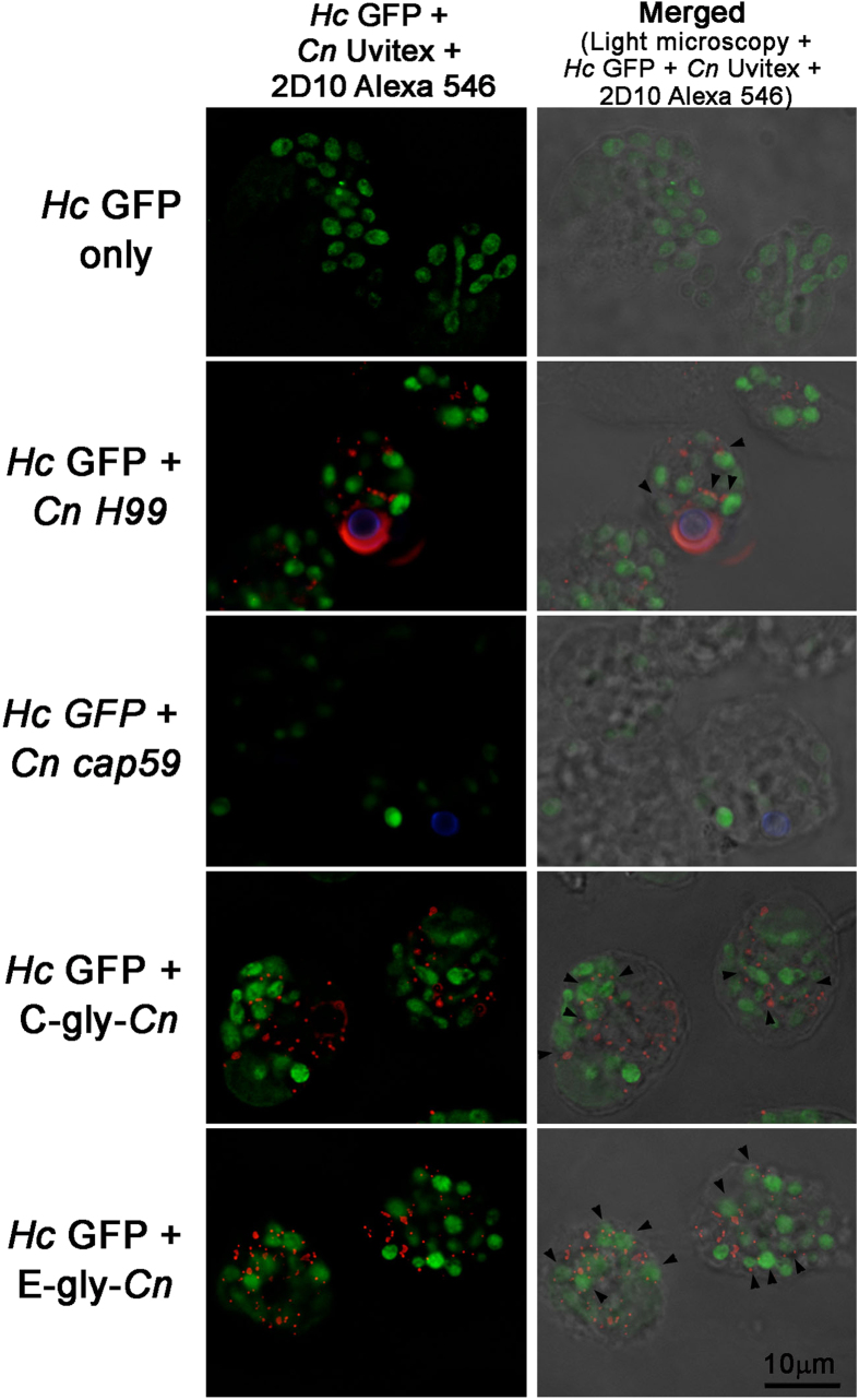
Figure 9. Hc co-localize with Cn-gly and is able to incorporate these glycans on its surface within the macrophage environment.
Macrophages were infected with Hc GFP (green) and incubated with either PBS, Cn H99 (Uvitex labeled – blue), Cn cap59 (Uvitex labeled – blue), C-gly-Cn or E-gly-Cn. Fluorescence was performed using 2D10 mAb and anti-IgM Alexa 568 conjugated (red). In the presence of Hc GFP yeasts and either Cn H99, C-gly-Cn or E-gly-Cn, Hc surface was labeled with 2D10 antibody as indicated in several instances by the black arrow heads. For Hc and PBS or Cn cap59 groups, no labelling for GXM was observed. Left column (Hc GFP-green; Cn Uvitex – blue; mAb 2D10 – red). Right column (Hc GFP-green; Cn Uvitex – blue; mAb 2D10 – red) merged with light microscopy. Scale bar = 10 μm.

