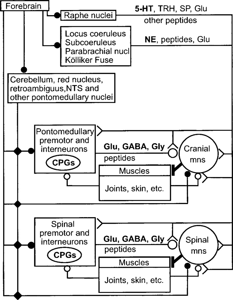
FIG. 4.
Anatomical organization of synaptic input to motoneurons. Main synaptic input to both cranial and spinal motoneurons comes from premotor and interneurons located close to the brain stem and spinal motoneuron pools; the few notable exceptions include direct corticospinal and rubrospinal inputs to motoneurons controlling the distal musculature, especially the digits, vestibulospinal projections to postural muscles, and bulbospinal projections transmitting inspiratory drive to phrenic motoneurons. Several cranial and spinal central pattern generators (CPG) are embedded in these premotor systems. The local premotor and interneurons also form the main gateway for relaying and integrating multisensorial afferent input from muscle, joints, skin, and descending synaptic information from forebrain, cerebellum, some brain stem nuclei, and the raphe, locus coeruleus, and other pontine/brain stem regions. Long projections from brain stem and pontine nuclei, both from diffusely projecting premotor groups, e.g., raphe and locus coeruleus, and from premotor groups involved in specialized motor tasks, e.g., respiration, equilibrium, posture, project directly to motoneurons, and to the local premotor and interneurons. Glutamate, GABA, and glycine are the principal transmitters of local premotor and interneurons but are also used in certain brain stem/pontine projections. 5-Hydroxytryptamine (5-HT), norepinephrine (NE), thyrotropin-releasing hormone (TRH), substance P (SP), and a host of other peptides are the main transmitters in the projections originating in brain stem/pontine nuclei, subserving modulatory functions in control of motoneuronal excitability. Symbols (solid circle, small open circle, large open circle, and fork shape) indicate different anatomical projection systems. Mns, motoneurons.

