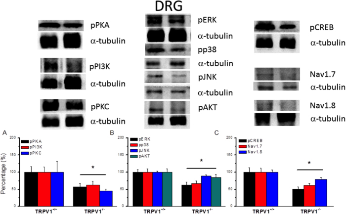
Figure 6. Expression levels of TRPV1 associated signaling pathways in L3–L5 DRG from TRPV1 null mice.
(A) Western blots of DRG lysates probed for pPKA, pPI3K, and pPKC. (B) Expression levels of pERK, pp38, pJNK, and pAKT. (A) Western blots of DRG lysates probed for pCREB, Nav1.7, and Nav1.8. α-tubulin was the internal control. *p < 0.05.

