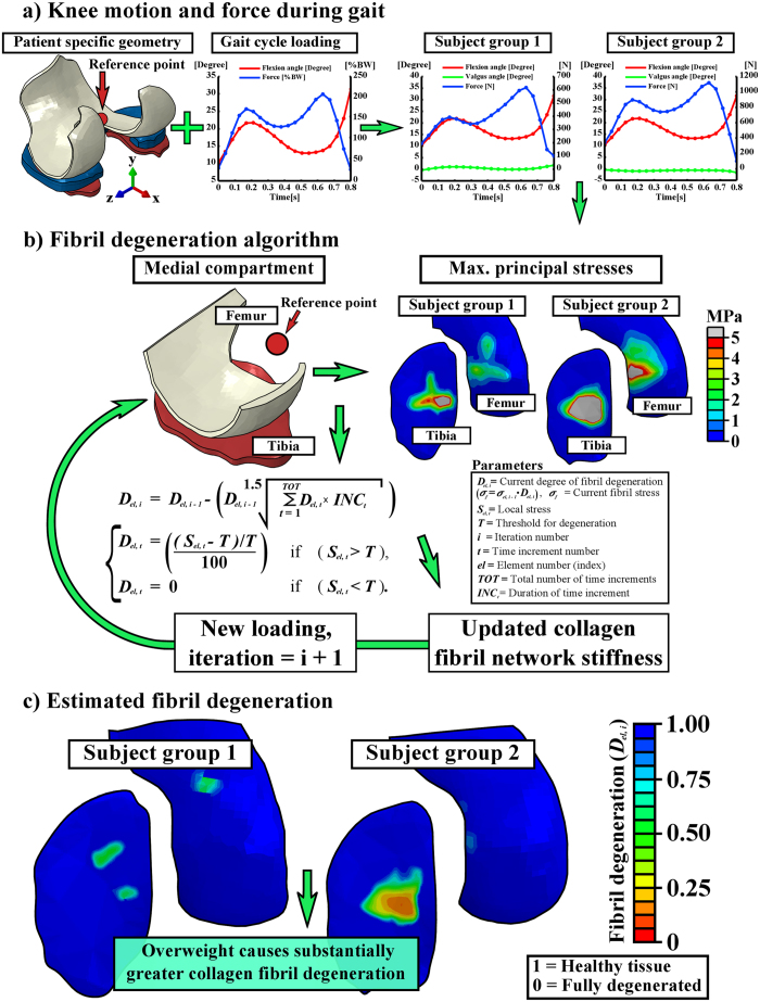
Figure 3.
Workflow from the subject-specific knee geometry with gait loading (a) to the fibril degeneration algorithm (b) and the results after iterative degenerative simulations (c). (a-left) Knee joint loads obtained from the literature24,28 were implemented into the subject-specific whole knee joint models. (a-right) The outcomes (force and rotations) of the whole knee joint model were implemented into the medial compartment model. (b) The fibril degeneration algorithm (left), based on excessive stresses (right), was applied into the medial compartment of the knee joint (most of the experimentally observed degeneration occurred there). The shown stress distributions are from the first peak loading force of the stance phase of gait. (c) Subject-specific fibril degenerations for both subject groups (healthy and obese). Used joint forces and rotations were considered as relative motions of femur with respect to tibia and they were implemented into a reference point located at the middle-central point between the medial and lateral epicondyles of the femur (a) (see details from Methods). In medial compartment models, (b) the location of the reference point was identical with that in the whole knee joint models (a).

