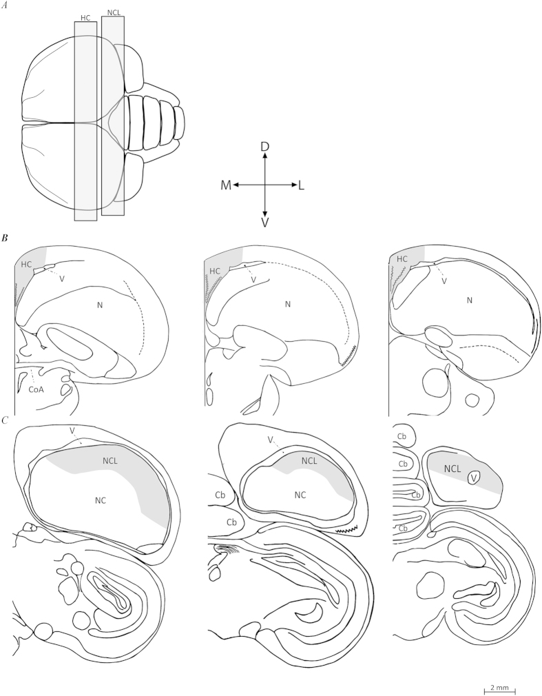
Figure 6. Schematic views of the two investigated brain regions in brains of turtle doves (Streptopelia turtur).
(A) Top view of the brain: rostral is to the left, caudal is to the right. We indicate the range within which frontal sections were taken from the hippocampal complex (HC), and nidopallium caudolateral (NCL). Seven sections were sampled along the rostro-caudal axis of each brain region (for details, see text), three of which are shown here: the most rostral, the middle, and the most caudal (from left to right), in HC (B), and NCL (C). Abbreviations: Cerebellum (Cb), Commissura anterior (CoA), Nidopallium (N), Nidopallium caudale (NC), Lateral ventricle (V). Orientations: Dorsal (D), Lateral (L), Ventral (V), and Medial (M). Created from images originally appearing in: Karten, Harvey J., and William Hodos57. A Stereotaxic Atlas of the Brain of the Pigeon (Columbia Livia). pp. 47-54, 61-67. © 1967 The Johns Hopkins Press. Adapted and reprinted with permission of Johns Hopkins University Press.

