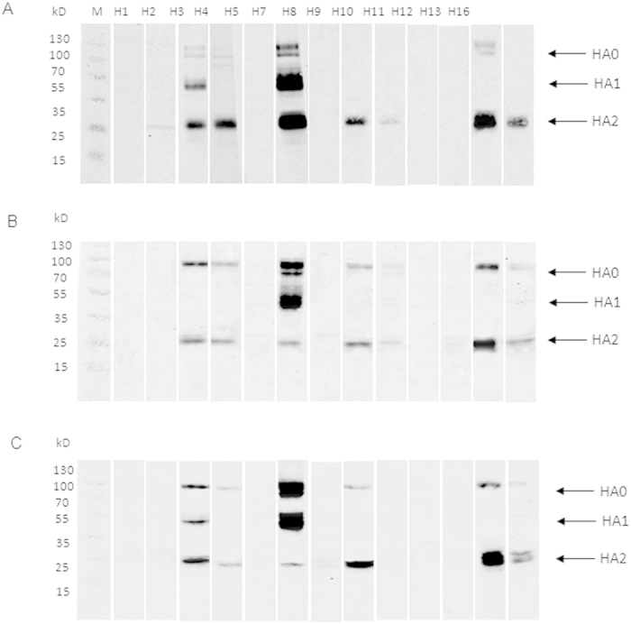
Figure 5. Identification of the cross-reactive regions by Western blot.

The lysates of MDCK cells infected with Anhui/1 (A), Shanghai/1 (B), and Shanghai/2 (C) isolates at an MOI of 0.1 were harvested 48 h post infection and probed with antisera to HA proteins of H1, H2, H3, H4, H5, H8, H9, H10, H11, H12, H13, and H16. Antisera against H7N9 HA were used as positive control.
