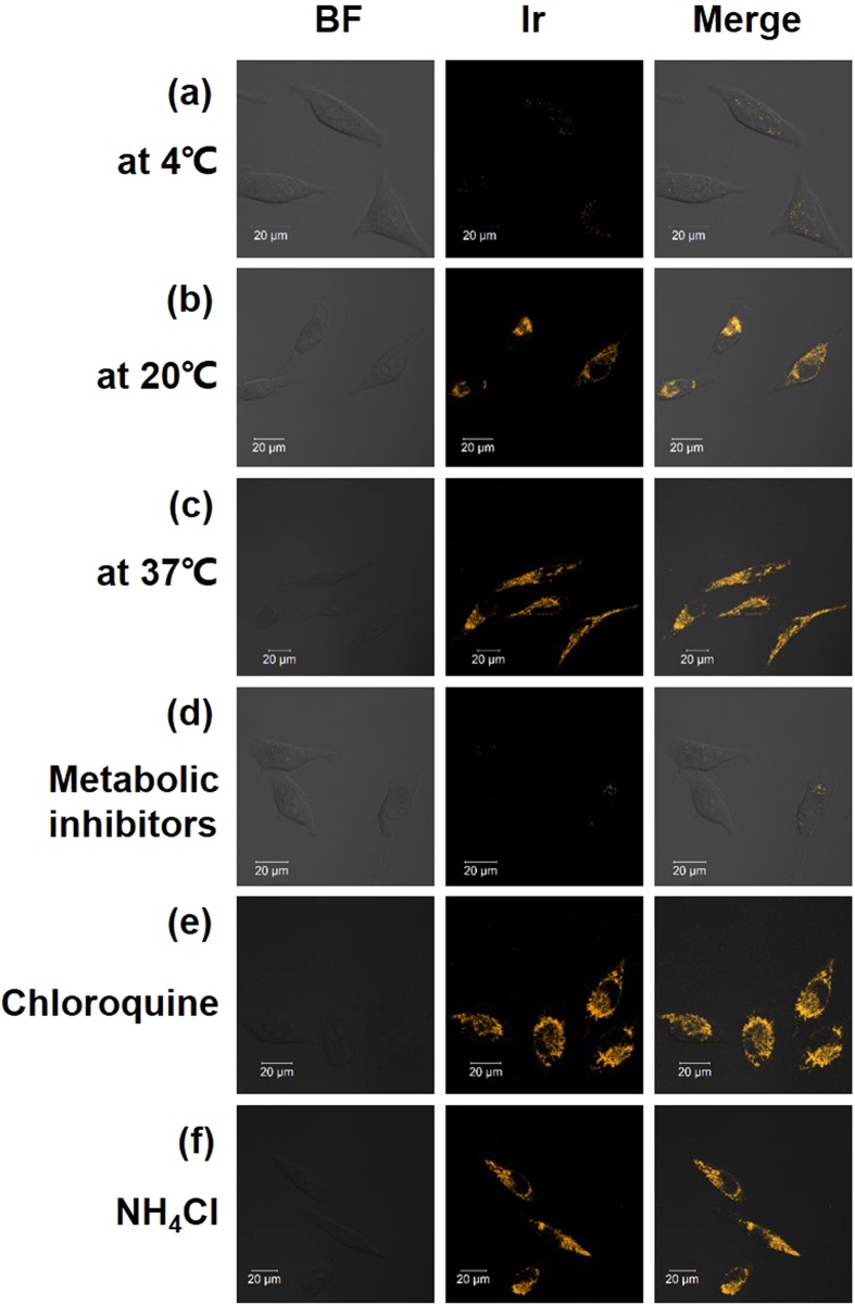
Figure 6. Confocal luminescence image and bright-field images of living HeLa cells incubated with 500 nM Ir1 in DMSO–PBS (pH 7.4, 1: 50, v/v) under different conditions.
(a–c) The cells were incubated with 500 nM Ir1 at 4 °C, 20 °C and 37 °C for 8 min, respectively. (d) The cells were preincubated with 50 mM 2-deoxy-D-glucose and 5 μM oligomycin in PBS for 1 h at 37 °C and then incubated with 500 nM Ir1 at 37 °C for 8 min. (e,f) The cells were pretreated with endocytic inhibitors (chloroquine (50 μM) and NH4Cl (50 mM), respectively) and then incubated with 500 nM Ir1 at 37 °C for 8 min (λex = 405 nm, λem = 590 ± 30 nm).

