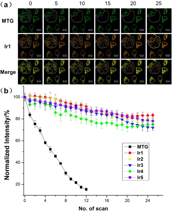
Figure 7. Photobleaching experiments of the Ir(III) complexes in HeLa cells.
(a) Time-dependent confocal imaging of Ir1/MTG co-stained HeLa cells. Confocal images of HeLa cells stained with Ir1 and MTG before and after verified scans of light irradiation. Time interval per scan: 15 s. Scale bar: 20 mm (λex = 405 nm, λem = 590 ± 30 nm). (b) Quantitative photobleaching results indicate that Ir1–Ir5 exhibited robust emission intensity under continuous light irradiation.

