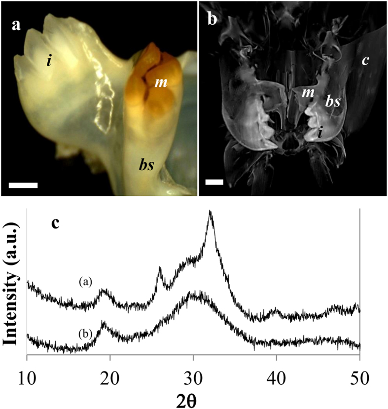
Figure 4. Mandible of the prawn, M. rosenbergii.
(a) Micrographs of the teeth processes illustrate the reverse distribution of chitin coat in the mandible compared to crayfish. In M. rosenbergii, the incisors are highly mineralized while the molar is less mineralized and is coated with chitin. (b) X-ray computer tomography of the mouth parts area demonstrates the gradual changes of mineral density from the teeth processes to the jaw and the rest of the head. The bulk cuticle is relatively lightly calcified with ACC while the mandibles show high mineral density, especially in the incisors (i-incisor, m-molar, bs-basal segment, c-cuticle, scale bar = 2 mm). (c) Powder XRD pattern of the molar (a) and the incisor (b) teeth. The diffractogram indicates that the molar tooth is composed of apatite while the incisor tooth is composed of ACP.

