Figure 7.
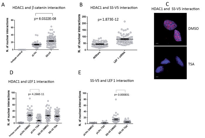
(A) SS18-SSX expression promotes interaction between β−catenin and HDAC1 in nuclei. Graphical representation with statistical analysis of PLAs using antibodies against the indicated proteins in C3H10T1/2pLIVc (pLIVc) and C3H10T1/2SS18-SSX1-V5 (SS-V5) cells. Correspondent representative fluorescence microscopy images are reported in Fig. S7. (B) LEF-1 depletion promotes SS18-SSX-HDAC1 association. Graphical representation of PLA as in (A). (C) TSA treatment inhibits SS18-SSX association with HDAC1. Representative fluorescence microscopy images of PLA using anti-V5 and anti-HDAC1 antibodies in C3H10T1/2SS18-SSX1-V5 cells stimulated with 250 nM TSA or DMSO for 16hrs. Scale bar: 5 μm. (D,E) Effect of TSA treatment on LEF-1 interactions with HDAC (D) and SS18-SSX (E); graphical representation of PLA as in (A).

