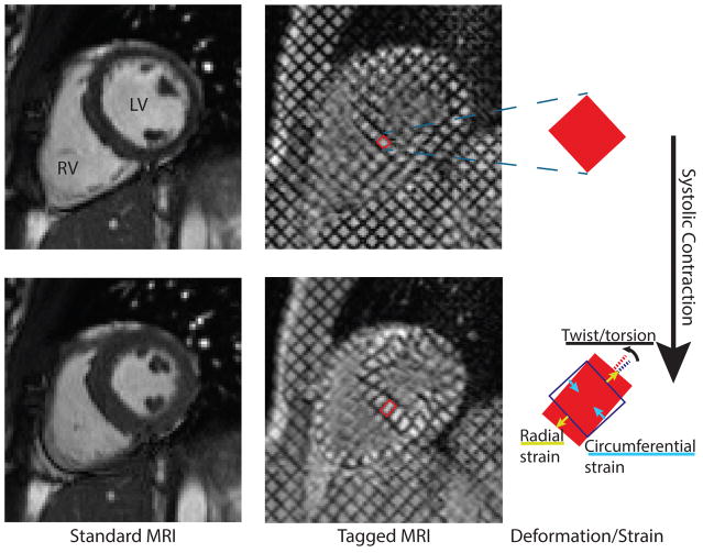
Fig. 1.
Normal heart contraction visualised with ‘standard’ and ‘tagged’ MRI. MR tagging uses spatial modulation of the magnetic field to superpose a grid pattern on the signal produced by the tissue at a given instant in time (usually end-diastole). The deformation of the heart muscle deforms the grid, so observing the deformation of the grid elements—or tags—provides a direct measurement of myocardial motion. This quantitative deformation data can be used to compute cardiac mechanics, such as radial or circumferential strain and torsion/twist

