
Fig. 3.
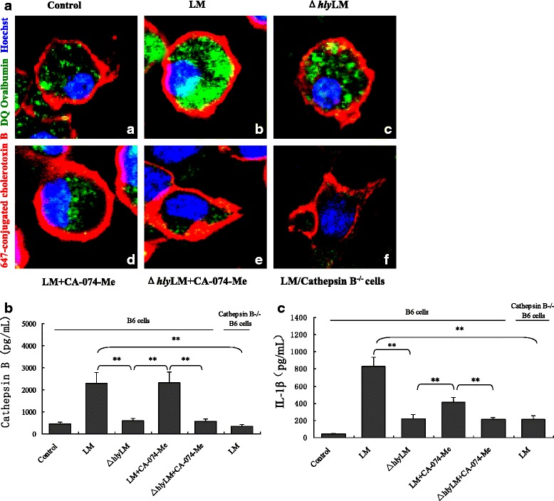
LM-induced lysosome damage and cathepsin B release from lysosomes in mouse B6 macrophage. a Confocal imaging of cathepsin B staining with DQ ovalbumin (green) in mouse B6 macrophages and cathepsin B−/− B6 macrophages infected with LM or Δhly LM (MOI = 20), with or without cathepsin B inhibitor CA-074-Me (The red color presented membrane and blue color presented nuclear): (a) untreated B6 cells; (b) LM-infected B6 cells; (c) Δhly LM-infected B6 cells; (d) and (e) LM- and Δhly LM-infected B6 cells pretreated with CA-074-Me (10 μM); (f) LM-infected cathepsin B−/− B6 cells. b and c cathepsin B in the cytosol and IL-1β in the culture medium of LM- and Δhly LM-infected B6 cells and cathepsin B−/− B6 cells following different treatments. The data in b and c are represented as means ± standard deviation from three independent experiments. * p < 0.05; ** p < 0.01. NS: no significance