
Fig. 3.
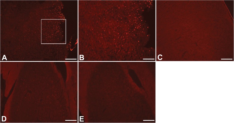
TUNEL staining of the cortex and hippocampus. TUNEL staining showed distribution of injured cells in the cortex similar to FJB as they were distributed throughout all layers of the cortex (a, b). However, no TUNEL staining was detected in the ipsilateral hippocampus (d). No TUNEL was observed on the contralateral side of the brain (C: cortex; E: hippocampus). TUNEL: red; Scale bars: 200 μm ( a , c - e ), 100 μm ( b )