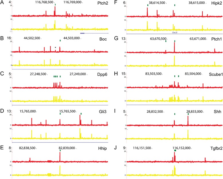
Fig. 5.

K-mer weights plotted across sequences that show enhancer activity. Diagrams were generated in UCSC Genome browser and show coordinate information for regions annotated to Ptch2 (a), Boc (b), Dpp6 (c), Gli3 (d), Hhip (e), Hipk2 (f), Ptch1 (g), Scube1 (h), Shh (i) and Tgfbr2 (j). Green boxes represent GBM. Weights for LDwGBM and NPwGBM are represented by the red and yellow lines, respectively. Refseq gene annotations are represented in blue. A putative Krox-20 TFBS (*) that has a high weight in the LDwGBM classifier but not the NPwGBM classifier occurs in the sequence annotated to Boc. Note that most sequences show weighted k-mers located several hundred bp from the central GBM, suggesting that sequence motifs that predict Hh enhancer activity may be distributed
