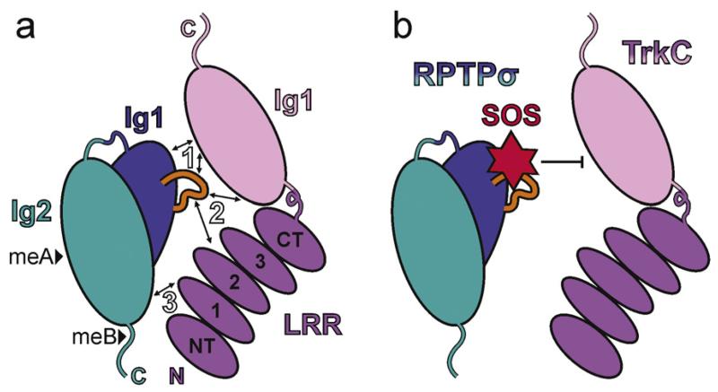
Fig. 4.
Synaptic type IIa RPTP-ligand complexes. (a) Cartoon illustrating the trans interaction mode observed in the chicken RPTPσ Ig12:TrkC LRRIg1 crystal structure (PDB accession code 4PBW) [57]. The three major binding sites are indicated by arrows and white labels. Protein features labelled as follows: Ig, immunoglobulin-like domain; LRR, leucine rich repeat domain (containing NT, amino-terminal cap; 1–3, leucine rich repeats; CT, carboxy-terminal cap); N, amino-terminus; C, carboxy-terminus; black arrowheads, potential meA and meB insertions; orange loop, RPTPσ Lys-loop. (b) Cartoon to illustrate the overlap of the sucrose octasulphate binding site on LAR (based on PDB accession code 2YD8 [58]) with the RPTPσ:TrkC interface. HS and heparin oligosaccharides have been shown to compete with TrkC for RPTPσ binding [57].

