
Fig. 1.
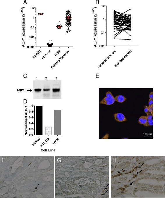
AQP1 expression. (a) relative expression of AQP1 (qPCR) in HUVEC, colon cancer cell lines, and human colon tumours (error bars show mean ± SEM; ** p = 0.004 ANOVA); (b) relative expression of AQP1 in CRC patients’ tumours compared to their matched normal mucosa. (c) western blot showing AQP1 monomer: lane 1 HUVEC, lane 2 HCT-116, lane 3 HT29. (d) corresponding quantification of bands using Image Lab™ Software (Bio-Rad); (e) immunofluorescence (IF) of HT29 colon cancer cells stained with anti-AQP1 and goat anti-rabbit secondary–Alexa 468 conjugate (orange) with NucBlue® stained nuclei overlay (63 x objective, scale bar = 10 μm). (f, g, h) immunohistochemistry of colon tumour sections staining for AQP1: F low, G moderate, H high expression. Arrows show examples of strong AQP1 staining of microvessels (20 x objective, scale bar = 0.1 mm)