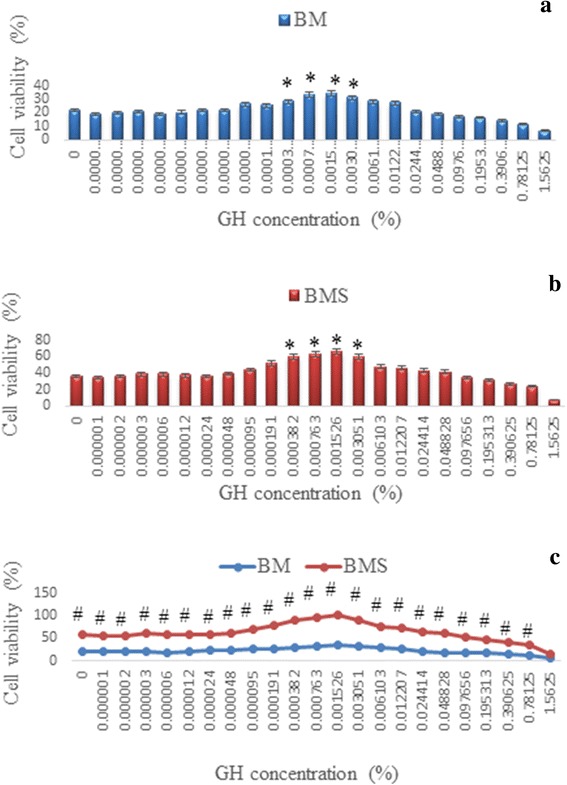
Fig. 1.

Corneal keratocytes proliferation in (a) Basal medium (BM), (b) Basal medium with serum (BMS), supplemented with serial dilution of GH and (c) comparison between the two media, BM and BMS. * denotes significant difference compare to control in the same group (p < 0.05), # denotes significant difference at the same concentration in different groups (p < 0.05)
