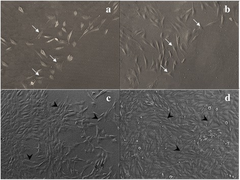
Fig. 2.

Phase contrast micrographs of corneal keratocytes cultured in four different media. a Basal medium (BM), (b) BM + 0.0015 % GH, (c) Basal medium with serum (BMS) and (d) BMS + 0.0015 % GH. Micrographs were taken at Passage 1 Day 3 of culture. Cell density was the least in BM medium (a) and the highest in BMS + 0.0015 % GH (d). Corneal keratocytes cultured in BM only (a) and with GH (b) exhibited shorter and broader cell morphology (thin arrow) compared to that of elongated and fibroblastic-shaped cells (thick arrow) in BMS medium (c) and with GH (d). Magnification (x50)
