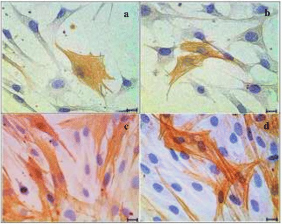
Fig. 7.

Immunocytochemistry of α-SMA in four different media. a Basal medium (BM), (b) BM + 0.0015 % GH, (c) Basal medium with serum (BMS) and (d) BMS + 0.0015 % GH. Positive stained cells showed stronger intensity in serum enriched media (c) and (d) compared to basal media (a) and (b). Scale bar represents 20 μm
