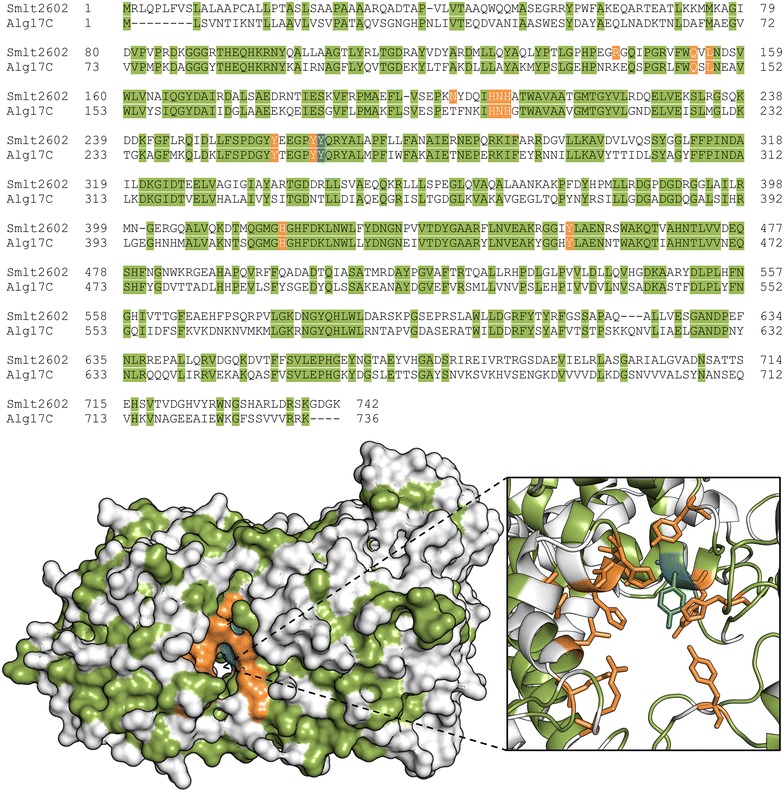
Fig. 2.

Identification of putative catalytic and substrate-binding residues in Smlt2602. Sequence alignment (top) and homology model (bottom) of Smlt2602 with S. degradans Alg17c lyase (Protein Data Bank 4OJZ) [13]. Identical residues are highlighted in green. The tyrosine residue predicted to act as the general acid in the β-elimination mechanism is highlighted in blue [13]. Residues predicted to be located near the active site cleft and to participate in either the catalytic mechanism or substrate binding are highlighted in orange
