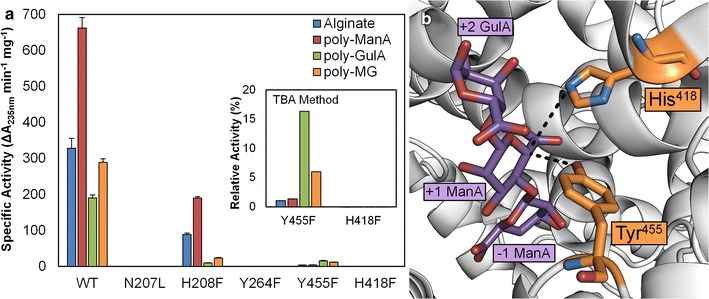
Fig. 3.

Mutagenesis of putative catalytic residues. a Specific activity of wild-type Smlt2602 and N207L, H208F, Y264F, H418F, and Y455F mutants against 1 mg/mL alginate-based substrates in 20 mM sodium phosphate buffer, pH 8.5. Enzyme activity was monitored by absorbance at 235 nm and confirmed via the TBA method (inset). For the TBA method, activity of WT against each substrate was taken to be 100 %. b Location of His418 and Tyr455 with respect to polysaccharide substrate, indicating each residue lies axial to the presumed location of the C5 proton (dashed black lines). Image generated from crystal structure of Alg17c from S. degradans 2–40 (Protein Data Bank 4OJZ) in complex with alginate trisaccharide (shown in purple). Residues in orange are His418 and Tyr450 of Alg17c which correspond to His418 and Tyr455 of Smlt2602 [13]
