
Figure 1.
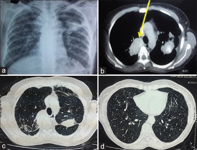
(Case 1): a) Chest radiograph showing bilateral reticulonodular opacities, upper lobe fibroparenchymal lesion and heterogenous opacity in left mid zone. b) CT chest showing partially calcified soft tissue mass in the right upper lobe 34×58mm (shown by arrow) and left upper lobe (35×51mm). c) Fibrotic changes seen in both upper lobes d) Randomly distributed nodules in bilateral lung