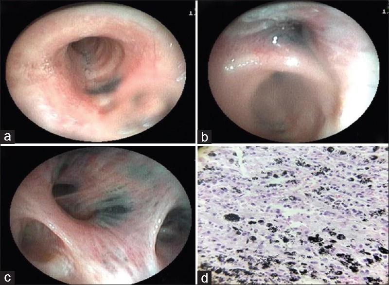
Figure 2.

(Case 1): (a and c) Bronchoscopy showing deposition of anthracotic pigments thought out bronchial tree on both sides and narrowing upper lobe bronchi due to b) external compression with distortion of bronchial lumen. d) Biopsy showing aggregates of polygonal cells with abundant black pigment in cytoplasm. Stroma was made up of fibrocollagenous tissue with neutrophils and mononuclear cells consistent diagnosis of anthracosis
