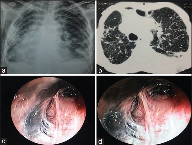
Figure 9.

(Case 6): a) Chest radiograph showing bilateral small nodular opacities in all zones with fibroparenchymal changes with pleural thickening. b) CECT chest showing randomly distributed nodules scatted in bilateral lung fields with fibroparenchymal changes and pleural reaction. (c and d) Bronchoscopy showing anthracotic pigments deposition throughout bronchial tree of both sides with luminal distortion
