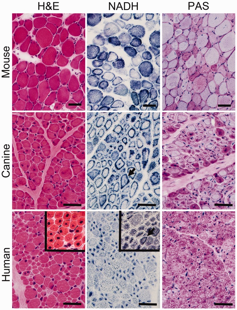
FIGURE 1.
Pathological findings in murine, canine, and human X-linked myotubular myopathy (XLMTM) by brightfield microscopy. Skeletal muscle from an Mtm1 knockout mouse at 43 days of life (mid-stage) are shown in comparison to an autopsy specimen from an 18-week-old (end-stage) canine with XLMTM and a diagnostic biopsy of a 2-month-old boy with XLMTM. Hematoxylin and eosin (H&E) staining reveals abnormal fiber size variation and the extent of central or internal nucleation in each affected species. In addition to the hypotrophy of some fibers seen at all disease stages, XLMTM shows marked atrophy of essentially all fibers in the terminal stage; therefore, the apparent difference in myofiber size between the murine (mid-stage) and canine models (end-stage) shown likely relates to disease stage. The percentage of centrally nucleated fibers can vary markedly among patients (H&E inset). Stains for reduced nicotinamide adenine dinucleotide (NADH, also known as NADH-TR) and periodic acid-Schiff (PAS) stain illustrate the patterns of organelle (mitochondrial/sarcotubular) and glycogen mislocalization, respectively, with pale peripheral halos in the human case but dark peripheral staining in the dog and mouse. Mouse tissue typically shows subsarcolemmal aggregation of mitochondria whereas true “necklace fibers” (arrows) can be seen in canine and some human (NADH inset) patient biopsies, as well as in rare fibers in the mouse. Scale bar = 40 µm.

