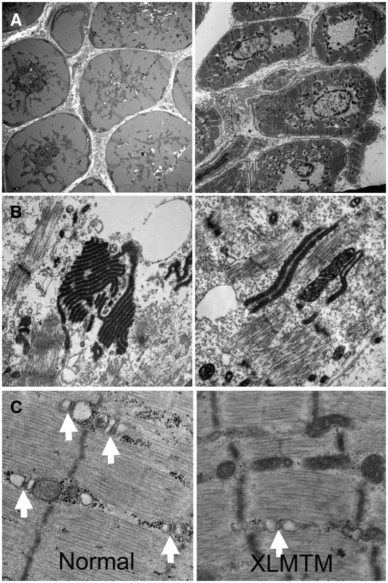
FIGURE 2.
Ultrastructural findings in human X-linked myotubular myopathy (XLMTM). (A) Low-power ultrastructural evaluations of XLMTM muscle reveal centrally located nuclei and central aggregates of organelles including mitochondria (left) and areas with only nuclei and glycogen (right). (B, C) Higher power shows organizational abnormalities of the sarcotubular system. Stacks of tubules (probably originating from triads) can be seen in a variety of configurations in the intermyofibrillar space, some of which contain osmiophilic material (B). There is also a lack of well-defined triad structures (white arrows) in XLMTM muscle in contrast to what is seen in normal muscle (C).

