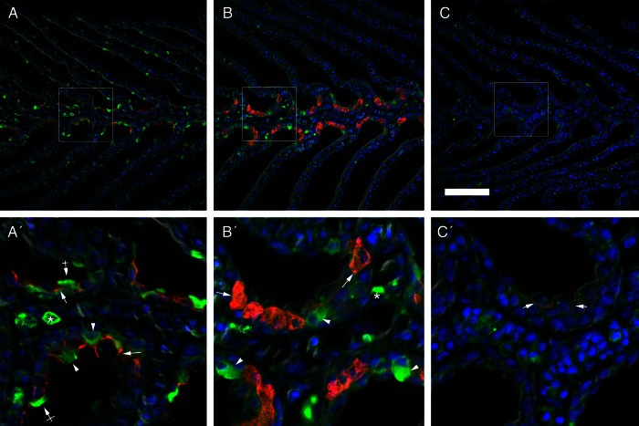
Figure 3:
Double immunofluorescence localization of V-ATPase (green) and Na+/K+-ATPase (red) with the corresponding merged image overlaid with DAPI nuclear staining (blue) in the gills of upstream-migrating lampreys in a freshwater-acclimated lamprey (A), a BW-25 osmoregulator (B) and a BW-25 osmocompromised animal (C). Arrows indicate NKA basolateral immunoreactivity. Arrowheads and crossed arrows indicate V-ATPase epithelial cytosolic and apical staining, respectively, while asterisks indicate leukocyte V-ATPase staining. Scale bar: 100 µm.

