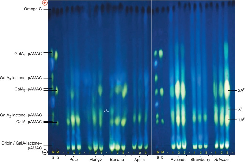
Fig. 3.
HVPE resolution of total Driselase digests of pAMAC-labelled AIR samples from seven fruit species. Fruit AIRs, each harvested at three stages of ripening (1–3; see Fig. 2), were successively treated with AMAC, acetone and Driselase (14 d); the pAMAC-labelled oligosaccharides generated were partially purified on a Supelco C18 cartridge column and de-lactonized in NaOH before electrophoresis. Each electrophoretogram loading was the products obtained from 20 mg f. wt of fruit tissue. Markers Ma and Mb are identical mixtures of acidic sugar–pAMAC conjugates before and after de-lactonization. Electrophoresis was at pH 6·5 and 4·0 kV for 45 min on Whatman No. 1 paper. Fluorescent spots were photographed under a 254-nm UV lamp. Orange G, loaded as a tracker between each fruit sample, shows up as a dark spot under UV. (+), anode; (–), cathode; –, blank loading.

