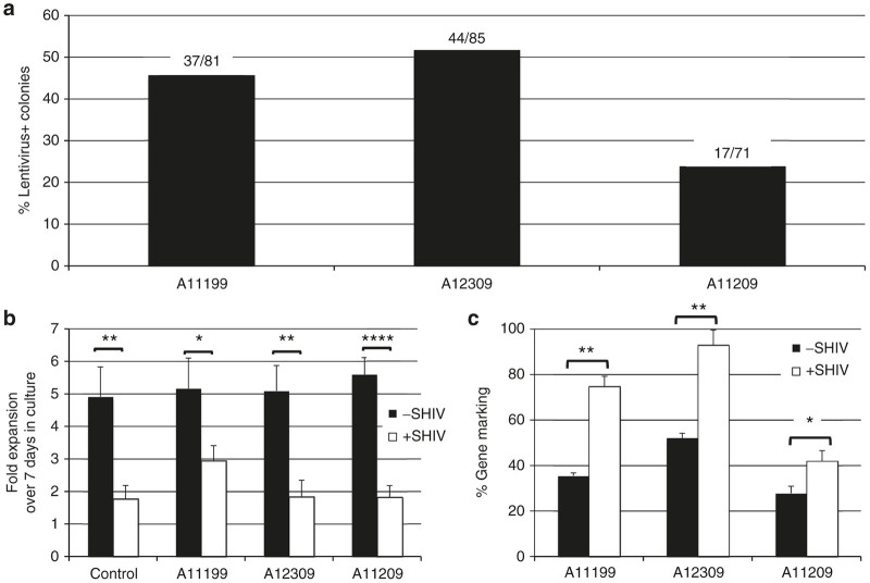
Figure 3.
Ex vivo measurements of Cal-1 gene marking and protection against SHIV challenge. (a) Prior to infusion into the indicated animal, Cal-1 GLP-transduced CD34+ cells were plated for colony forming assays. Colonies were picked from a total of three plates and screened by PCR for lentiviral backbone and actin. Values over each bar represent the number of lentivirus-positive colonies (numerator) as a function of total actin-positive colonies (denominator) (b, c). CD4+ cells were collected from the indicated animals following transplant recovery, challenged ex vivo with SHIV-Ku1 at a multiplicity of infection (MOI) of 0.05, and followed over a 7-day time course. b Fold expansion of cells from one non-transplanted control, and the indicated Cal-1-transplanted animals. c Lentivirus gene marking was measured by Taqman. Data represent average and standard error of the mean for at least three replicate analyses from two independent experiments. P values: *P ≤ 0.05; **P ≤ 0.01; ****P ≤ 0.0001. SHIV, simian/human immunodeficiency virus.

