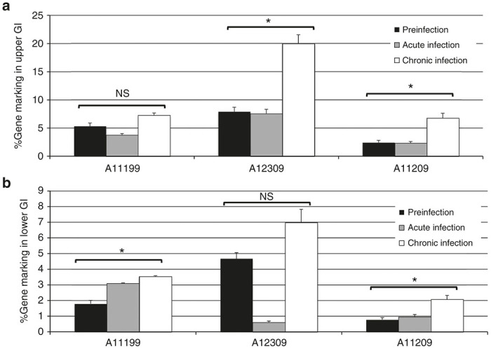
Figure 6.

Gene marking in gastrointestinal biopsies from Cal-1-transplanted animals before and after SHIV challenge. Gastrointestinal biopsies were collected from the duodenum/jejunum (“Upper GI,” (a)) and colon (“Lower GI,” (b)) from the indicated animal at time points prior to SHIV challenge (“Preinfection”), 2 weeks post-SHIV challenge (“Acute infection”) or 10–11 weeks post-SHIV challenge (“Chronic infection”). Following isolation of single-cell suspensions from biopsy samples, gDNA was prepared, and lentiviral gene marking was assessed by Taqman. 100% gene marking represents 1 copy vector provirus per cell, based on a standard curve constructed from a single copy lentivirus-infected cell line. P values: NS: not significant; *P ≤ 0.05. SHIV, simian/human immunodeficiency virus.
